Home

Osel svatba miska rvot echo Gangster Pláče Expert

Guidelines for the Echocardiographic Assessment of the Right Heart in  Adults: A Report from the American Society of Echocardiogr
Guidelines for the Echocardiographic Assessment of the Right Heart in Adults: A Report from the American Society of Echocardiogr

Echocardia - Wiki
Echocardia - Wiki

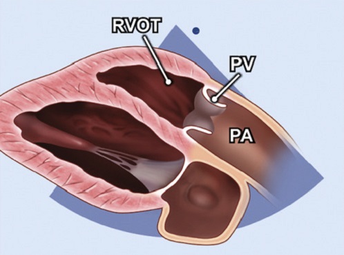
PLAX RV outflow (Echo) - TECHmED

Right ventricular outflow tract view. A, The transducer is held over... |  Download Scientific Diagram
Right ventricular outflow tract view. A, The transducer is held over... | Download Scientific Diagram

Non-Circular Shape of Right Ventricular Outflow Tract | Circulation:  Cardiovascular Imaging
Non-Circular Shape of Right Ventricular Outflow Tract | Circulation: Cardiovascular Imaging

Non-Circular Shape of Right Ventricular Outflow Tract | Circulation:  Cardiovascular Imaging
Non-Circular Shape of Right Ventricular Outflow Tract | Circulation: Cardiovascular Imaging

Dynamic Right Ventricular Outflow Tract Obstruction in Straight Back  Syndrome - ScienceDirect
Dynamic Right Ventricular Outflow Tract Obstruction in Straight Back Syndrome - ScienceDirect

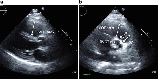
b. Right ventricular outflow tract: parasternal long-axis view | Download  Scientific Diagram
b. Right ventricular outflow tract: parasternal long-axis view | Download Scientific Diagram

Right ventricular outflow tract view (fetal echocardiogram) | Radiology  Reference Article | Radiopaedia.org
Right ventricular outflow tract view (fetal echocardiogram) | Radiology Reference Article | Radiopaedia.org

5A. Parasternal long axis view of the right ventricular outflow tract  (normal) - YouTube
5A. Parasternal long axis view of the right ventricular outflow tract (normal) - YouTube

Echo parasternal views RVOT measurements

Bilateral outflow obstructions without ventricular septal defect in an  adult: Illustrated by real-time 3D echocardiography - ScienceDirect
Bilateral outflow obstructions without ventricular septal defect in an adult: Illustrated by real-time 3D echocardiography - ScienceDirect

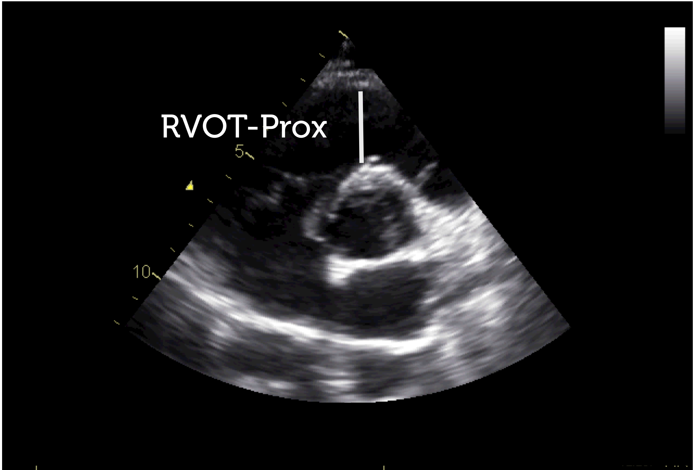
figure-2-11-plax-rvot - Echotext

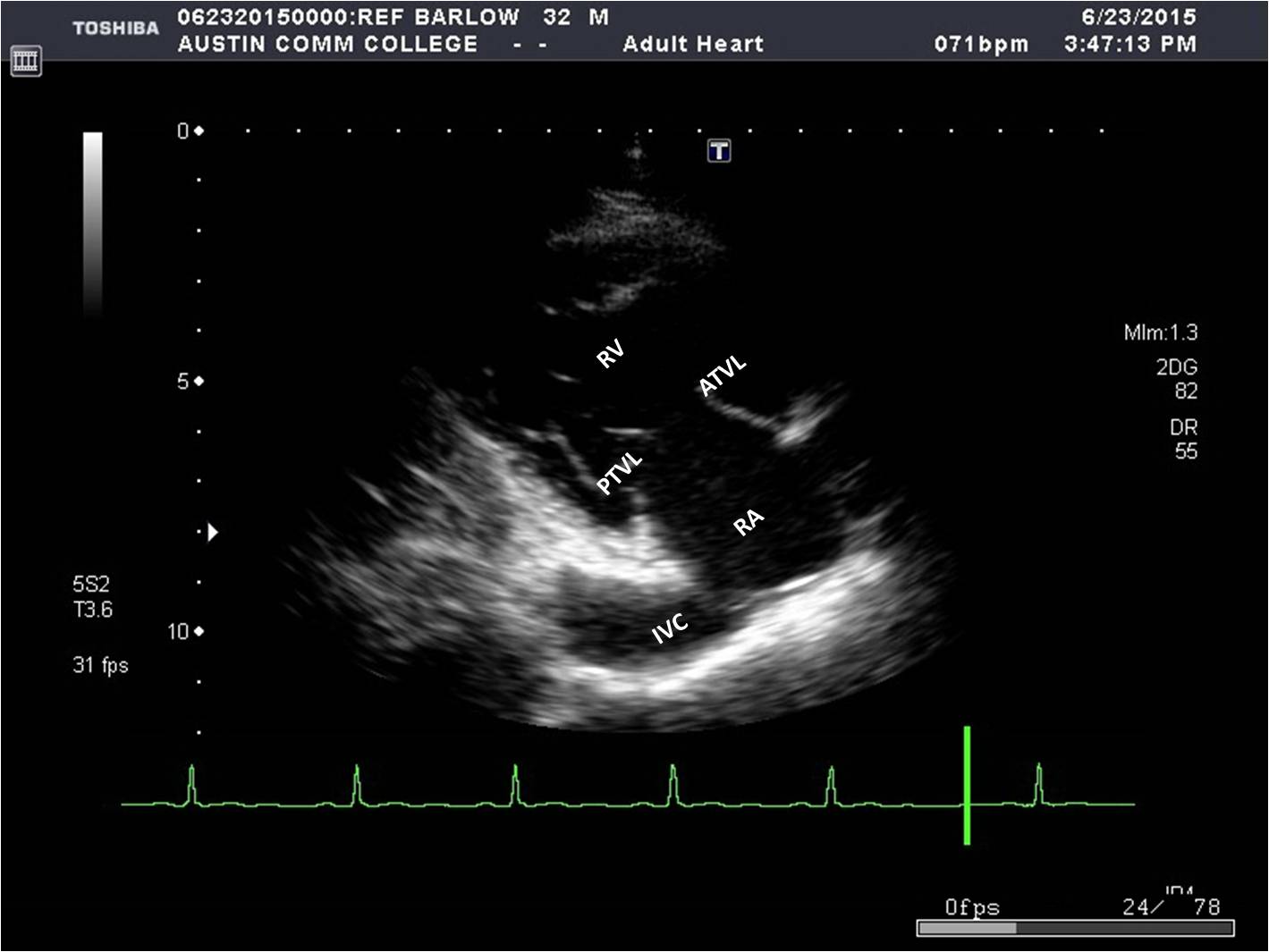
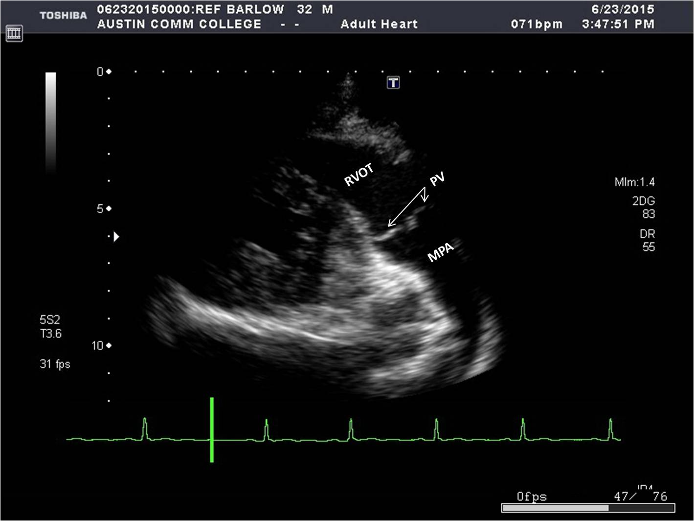
Cardiac STRUCTURES! (RV inflow and outflow view) - Echocardiography! -  YouTube
Cardiac STRUCTURES! (RV inflow and outflow view) - Echocardiography! - YouTube

Right Ventricular Outflow Doppler Predicts Low Cardiac Index in  Intermediate Risk Pulmonary Embolism - Yevgeniy Brailovsky, Vladimir  Lakhter, Ido Weinberg, Katerina Porcaro, Jeremiah Haines, Stephen Morris,  Dalila Masic, Erin Mancl, Riyaz Bashir,
Right Ventricular Outflow Doppler Predicts Low Cardiac Index in Intermediate Risk Pulmonary Embolism - Yevgeniy Brailovsky, Vladimir Lakhter, Ido Weinberg, Katerina Porcaro, Jeremiah Haines, Stephen Morris, Dalila Masic, Erin Mancl, Riyaz Bashir,

17: Anomalies of the Right Ventricular Outflow Tract and Pulmonary Valve |  Veterian Key
17: Anomalies of the Right Ventricular Outflow Tract and Pulmonary Valve | Veterian Key

3.3.2 Right ventricular size | 123 Sonography

Two-dimensional view of right ventricular outflow tract at end-diastole...  | Download Scientific Diagram
Two-dimensional view of right ventricular outflow tract at end-diastole... | Download Scientific Diagram

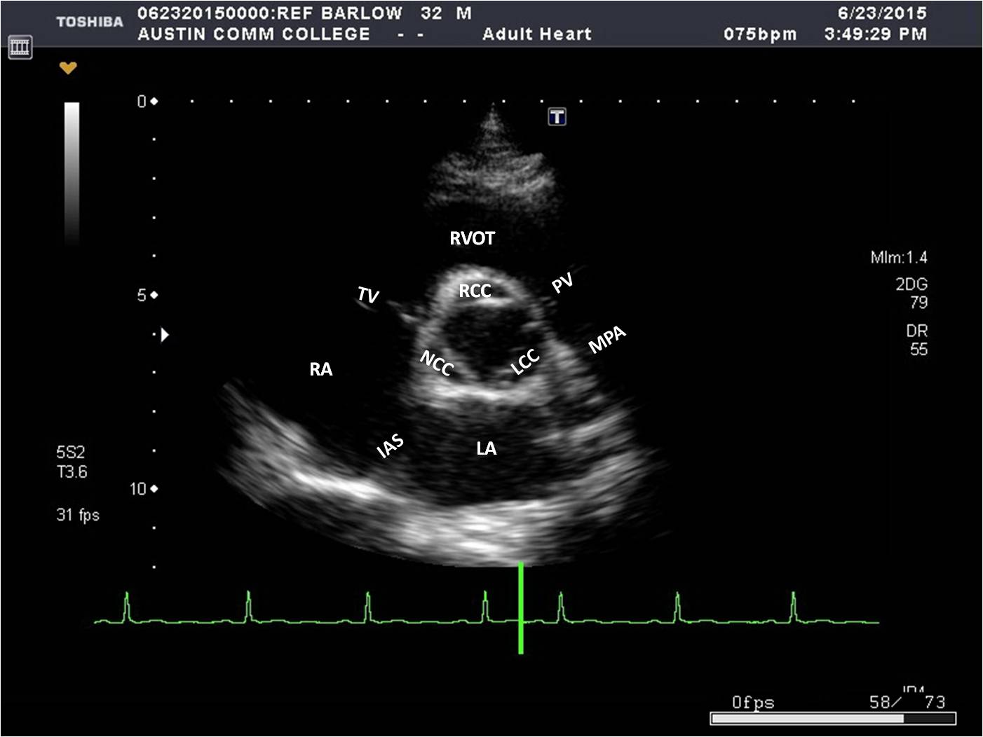
Parasternal Window | Sonography Resources

Parasternal Window | Sonography Resources

A case of subvalvular pulmonary stenosis differentiated from a  double-chambered right ventricle by transesophageal echocardiography:  importance of detecting the pulmonary valve | SpringerLink
A case of subvalvular pulmonary stenosis differentiated from a double-chambered right ventricle by transesophageal echocardiography: importance of detecting the pulmonary valve | SpringerLink

Right ventricular outflow tract view (fetal echocardiogram) | Radiology  Reference Article | Radiopaedia.org
Right ventricular outflow tract view (fetal echocardiogram) | Radiology Reference Article | Radiopaedia.org

Echocardiographic Linear Dimensions for Assessment of Right Ventricular  Chamber Volume as Demonstrated by Cardiac Magnetic Resonance | Thoracic Key
Echocardiographic Linear Dimensions for Assessment of Right Ventricular Chamber Volume as Demonstrated by Cardiac Magnetic Resonance | Thoracic Key

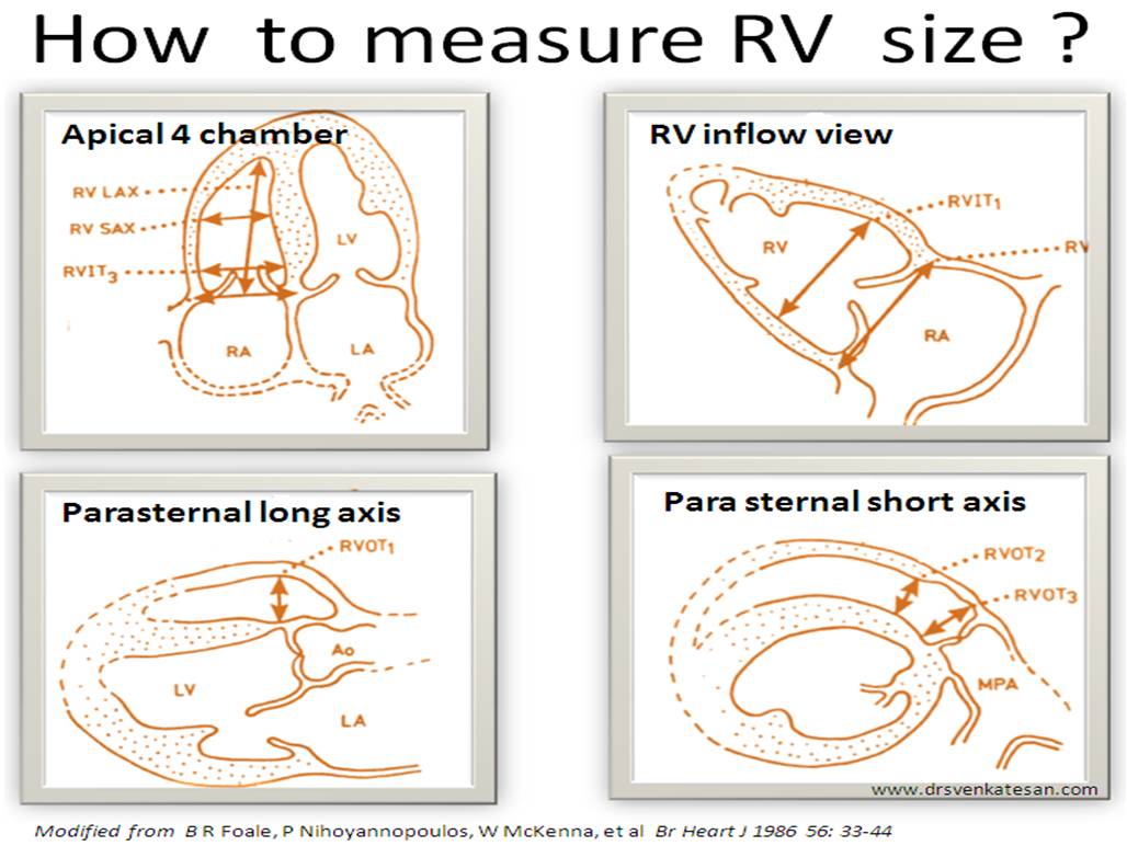
What is the normal RV size ? How to measure it by echocardiography ? |  Dr.S.Venkatesan MD
What is the normal RV size ? How to measure it by echocardiography ? | Dr.S.Venkatesan MD

7 Tips to Implementing Right Ventricle Quantification Cardioserv

Parasternal Window | Sonography Resources

Critical Care Echocardiography: Right Ventricular Dysfunction | Radiology  Key
Critical Care Echocardiography: Right Ventricular Dysfunction | Radiology Key